Arteries In Neck - Major Arteries of the Head and Neck - Medical Stock Images ...
Arteries In Neck - Major Arteries of the Head and Neck - Medical Stock Images .... These blood vessels can have abnormal shapes, sizes or paths through the neck and head. They are the carotid arteries, and they carry blood to the brain. This is called carotid artery disease, which increases your risk of stroke. The arteries in neck that supply blood to the brain are called carotid arteries. The carotid arteries provide the head's blood supply and run along both sides of the neck.
ads/bitcoin1.txt
The blockage increases your risk of stroke, a medical emergency that occurs when the blood supply to the brain is interrupted or seriously reduced. The carotid arteries are major blood vessels in the neck that supply blood to the brain, neck, and face. Two large arteries flow from the heart up the sides of the neck and into the brain. If one of them is narrowed or blocked, it can lead to a stroke. Doctors can test for a narrowed carotid artery, but it's usually not a good idea.
This diagram shows the veins present in the head and neck ... from i.pinimg.com The exam generally includes listening for a swooshing sound (bruit) over the carotid artery in your neck, a sound that's characteristic of a narrowed artery. When your doctor puts their hands on your neck to detect. The carotid arteries are major blood vessels in the neck that supply blood to the brain, neck, and face. Without this blood flow, your brain cells would. The plaque buildup is made of fat, cholesterol, cellular waste, calcium, proteins and inflammatory cells. A buildup of plaque can narrow or block your carotid arteries. While the stent is being placed, blood flow through the carotid artery is reversed temporarily. Two large arteries flow from the heart up the sides of the neck and into the brain.
Cervical in the neck, petrous in the base of the skull, cavernous within the cavernous sinus and intracranial above the cavernous sinus.
ads/bitcoin2.txt
Two large arteries flow from the heart up the sides of the neck and into the brain. A new classification system divides the internal carotid artery into four parts; Cervical in the neck, petrous in the base of the skull, cavernous within the cavernous sinus and intracranial above the cavernous sinus. When your doctor puts their hands on your neck to detect. The vertebral arteries stem from the subclavian arteries; You have two carotid arteries, one on each side of your neck. The internal carotid artery is one of two branches of the common carotid artery. It is responsible for supplying a large portion of the anterior and middle parts of the brain. The carotid artery brings needed blood to your brain and face. In the neck, the carotid sheath (fibrous connective tissue) covers the common carotid artery, vagus nerve, and internal jugular vein. The blockage increases your risk of stroke, a medical emergency that occurs when the blood supply to the brain is interrupted or seriously reduced. Though more often occurring with carotid arteries (the other major ones supplying the brain through the neck), vertebral arteries can be impacted. There are two carotid arteries, one on the right and one on the left.
Though more often occurring with carotid arteries (the other major ones supplying the brain through the neck), vertebral arteries can be impacted. In the neck, the carotid sheath (fibrous connective tissue) covers the common carotid artery, vagus nerve, and internal jugular vein. Your carotid arteries are the major blood vessels that deliver blood to your brain. However, pain from carotidynia typically only occurs on one side. The arteries in neck that supply blood to the brain are called carotid arteries.
Carotid Stenosis Treatments from fthmb.tqn.com A buildup of plaque can narrow or block your carotid arteries. Related posts of arteries in the neck picture veins and arteries of the neck. The arteries in the chest, neck and brain are the most frequent arteries found to be abnormal in phace syndrome. When your doctor puts their hands on your neck to detect. A new classification system divides the internal carotid artery into four parts; Though more often occurring with carotid arteries (the other major ones supplying the brain through the neck), vertebral arteries can be impacted. Two large arteries flow from the heart up the sides of the neck and into the brain. The carotid artery brings needed blood to your brain and face.
Blood is carried to the brain through blood vessels called arteries.
ads/bitcoin2.txt
Your carotid arteries are the major blood vessels that deliver blood to your brain. Veins and arteries of the neck 9 photos of the veins and arteries of the neck activate javascript arteries in the neck diagram, common carotid artery branches, external carotid artery function, how many carotid arteries, left common carotid artery function, the left common carotid artery supplies blood to the. Two large arteries flow from the heart up the sides of the neck and into the brain. It is responsible for supplying a large portion of the anterior and middle parts of the brain. The arteries in the chest, neck and brain are the most frequent arteries found to be abnormal in phace syndrome. It involves making a tiny incision at the base of the neck and, from there, inserting a stent into the carotid artery. You have two carotid arteries, one on each side of your neck. The left and right common carotid arteries ascend up the neck, lateral to the trachea and the oesophagus. One carotid artery is located on each side of your neck. Blood is carried to the brain through blood vessels called arteries. While the stent is being placed, blood flow through the carotid artery is reversed temporarily. However, pain from carotidynia typically only occurs on one side. They are the carotid arteries, and they carry blood to the brain.
It is responsible for supplying a large portion of the anterior and middle parts of the brain. The exam generally includes listening for a swooshing sound (bruit) over the carotid artery in your neck, a sound that's characteristic of a narrowed artery. It involves making a tiny incision at the base of the neck and, from there, inserting a stent into the carotid artery. Cervical in the neck, petrous in the base of the skull, cavernous within the cavernous sinus and intracranial above the cavernous sinus. However, neck arteries can work just as fine, even though they are partially blocked.
Subclavian Artery Photographs | Fine Art America from render.fineartamerica.com Two large arteries flow from the heart up the sides of the neck and into the brain. Cadaveric angiographic and dissection studies have demonstrated that the external and internal carotids are the main arterial sources for the head and… Blood flow in this artery can become partly or totally blocked by fatty material called plaque. It involves making a tiny incision at the base of the neck and, from there, inserting a stent into the carotid artery. However, pain from carotidynia typically only occurs on one side. You have one of these arteries on each side of your neck. Though more often occurring with carotid arteries (the other major ones supplying the brain through the neck), vertebral arteries can be impacted. The blockage increases your risk of stroke, a medical emergency that occurs when the blood supply to the brain is interrupted or seriously reduced.
They are the carotid arteries, and they carry blood to the brain.
ads/bitcoin2.txt
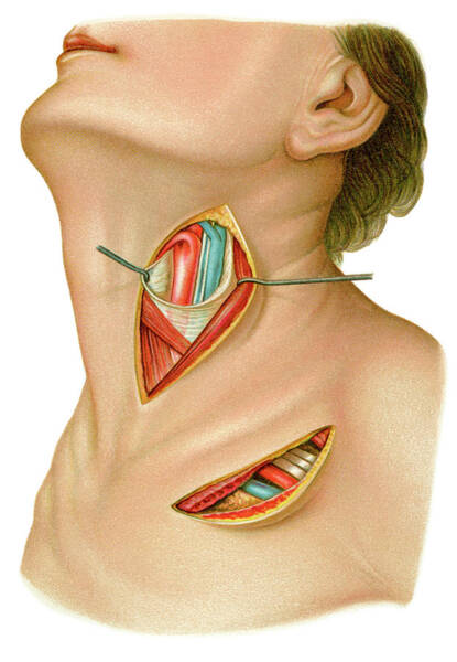
Two pairs of blood vessels in the neck — the carotid and vertebral arteries, known collectively as the cervical arteries — carry blood to the brain. The carotid arteries are two large blood vessels that supply oxygenated blood to the large, front part of the brain. Without this blood flow, your brain cells would. It involves making a tiny incision at the base of the neck and, from there, inserting a stent into the carotid artery. A new classification system divides the internal carotid artery into four parts; Veins and arteries of the neck 9 photos of the veins and arteries of the neck activate javascript arteries in the neck diagram, common carotid artery branches, external carotid artery function, how many carotid arteries, left common carotid artery function, the left common carotid artery supplies blood to the. At the root of the neck the right internal jugular vein is placed at a little distance from the common carotid artery, and crosses the first part of the subclavian artery, while the left internal jugular vein usually overlaps the common carotid artery. There are two carotid arteries, one on the right and one on the left. Just like other arteries in the body, neck arteries are also susceptible to blockages. A tear in the lining of one of these vessels is called a cervical artery dissection. While the stent is being placed, blood flow through the carotid artery is reversed temporarily. When your doctor puts their hands on your neck to detect. In the neck, the carotid sheath (fibrous connective tissue) covers the common carotid artery, vagus nerve, and internal jugular vein.
ads/bitcoin3.txt
ads/bitcoin4.txt
ads/bitcoin5.txt
0 Response to "Arteries In Neck - Major Arteries of the Head and Neck - Medical Stock Images ..."

/GettyImages-548001367-573f60493df78c6bb015ec20.jpg)

0 Response to "Arteries In Neck - Major Arteries of the Head and Neck - Medical Stock Images ..."
Post a Comment